FIG. 8.
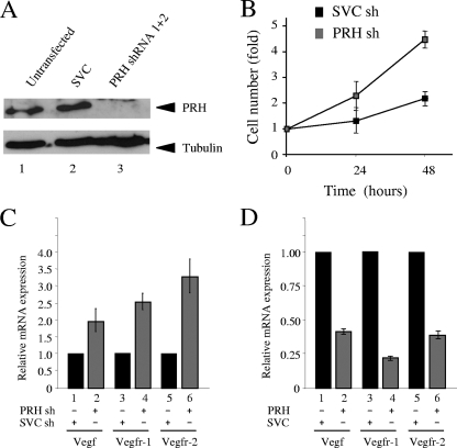
PRH represses VSP genes in MCF-7 cells. (A) Western analysis of whole-cell extracts from untransfected MCF-7 cells or cells cotransfected with SVC shRNA or PRH shRNA (1+2). Extracts were stained with rabbit PRH antisera (top) and tubulin antibody (bottom). (B) Growth curves for MCF-7 cells transfected with SVC shRNA (sh) or PRH shRNA (1+2). Cells were selected with puromycin 24 h posttransfection, and after 5 days in selection, MTT assays were performed over 3 days. Results shown are representative of the results from 6 independent experiments. (C) Vegf, Vegfr-1, and Vegfr-2 mRNA levels in MCF-7 cells after shRNA cotransfection (as above). Levels of mRNA were determined by qRT-PCR using specific primers and compared to that for GAPDH. Black bars represent the SVC shRNA-targeted cells and gray bars the PRH shRNA (1+2)-targeted cells. Values are means and SD (n = 5). (D) Vegfr-1, Vegfr-2, and Vegf mRNA levels in MCF-7 cells 48 h after transfection with pMUG1 (empty vector) or pMUG1-Myc-PRH. mRNA levels were determined as above. Values are means and SD (n = 3).

