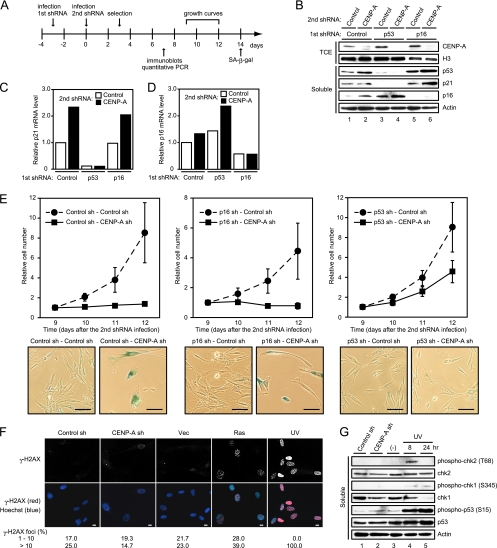
FIG. 7.
Senescencelike phenotypes induced by CENP-A depletion require p53. (A) Experimental design. (B) Immunoblotting of TIG3 cells expressing control, p53, or p16INK4a shRNA (first shRNA) in combination with control or CENP-A shRNA (second shRNA). TCE and soluble proteins were resolved by SDS-PAGE, followed by immunoblotting with the indicated antibodies. Histone H3 and actin were used as loading controls. (C and D) Quantitative RT-PCR of the cells indicated in panel B. The relative abundances of p21CIP1 mRNA (C) and p16INK4a mRNA (D) are shown after normalization using GAPDH mRNA. (E) Growth curves of TIG3 cells expressing control (left upper), p16INK4a (middle upper), or p53 shRNA (right upper) in combination with control or CENP-A shRNA. The number of cells on day 9 after the second shRNA infection was set at 1. The data are means ± the SD from three independent experiments. SA-β-Gal staining was performed on day 14 after the second shRNA infection (lower panels). Scale bars, 100 μm. (F) Immunofluorescence of CENP-A-depleted and ras-induced senescent TIG3 cells performed on day 10 after infection. Staining revealed γ-H2AX (red in overlay). DNA was counterstained with Hoechst (blue in overlay). As a positive control, cells were treated with UV. The numbers of cells (n = 300) containing 1 to 10 and >10 foci are shown. Scale bars, 10 μm. (G) Immunoblotting of CENP-A-depleted and UV-treated cells. Soluble proteins were resolved by SDS-PAGE, followed by immunoblotting with the indicated antibodies. Actin was used as a loading control.

