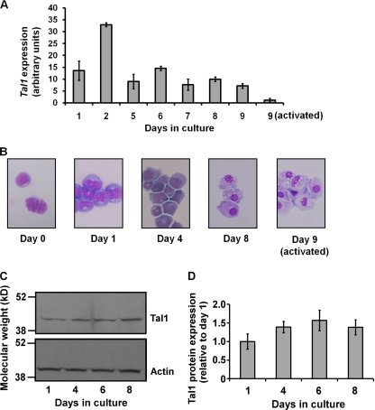
FIG. 1.
Tal1 mRNA expression during in vitro differentiation of CMPs to macrophages and following LPS/IFN-γ activation. (A) Total RNAs prepared from purified precursors and differentiated cells at the indicated times in culture were converted to cDNA for real-time PCR analysis employing SYBR green fluorescence. Each bar represents the mean ± the standard deviation from three independent PCRs as normalized to RPS16 mRNA expression. Results are representative of three independent experiments. (B) Morphology of differentiating cells from Wright-Giemsa stains of cytospin preparations. (C) Western blot analysis of Tal1 expression in cultures of terminally differentiating mouse BM MM precursors. Results are representative of two independent analyses. (D) Graph of normalized Tal1 expression from Western blot analysis.

