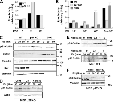
FIG. 3.
Rho A activity is high in p27 KO cells following adhesion to ECM substrates. (A) Results of a pulldown assay of active RhoA in WT and p27 KO MEFs incubated in reduced serum (0.1%) medium overnight and then stimulated with bFGF (25 ng/ml) for the indicated times. (B) Results of a pulldown assay of active RhoA in WT, p27 KO, and DKO MEFs. Cells were serum starved for 24 h, detached, and kept in suspension for 30 min (Sus) and then adhered to FN (30 μg/ml) for the indicated times. Data represent the means of three independent experiments. (C) Western blot analysis of pSer3-cofilin on lysates from WT, p27 KO, and DKO MEFs alowed to adhere to FN (10 μg/ml) for the indicated times. Standard molecular weights are indicated on the right. Vinculin was used as a loading control. (D) Western blot analysis of pSer3-cofilin on lysates from p27 KO MEFs allowed to adhere to FN (10 μg/ml) for the indicated times in the presence of C3-exoenzyme (2 ng/ml) or Y27632 (10 μM). (E) Western blot analysis of pSer3-cofilin on lysates from WT MEFs treated with increasing doses of nocodazole (upper panel) or cells allowed to adhere to FN (10 μg/ml) for the indicated times in the presence of nocodazole at the indicated doses (lower panel). Standard molecular weights are indicated on the right. (F) Western blot analysis of pSer3-cofilin in lysates from p27 KO MEFs allowed to adhere to FN (10 μg/ml) for the indicated times in the presence of 100 nM paclitaxel. Standard molecular weights are indicated on the right.

