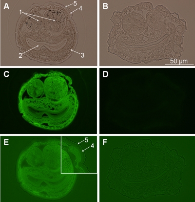
FIG. 2.
Transverse sections through adult Haemonchus contortus probed with fluorescein-conjugated avidin. Sections of adult nematodes following incubation with a biotinylated form of [N29K]-kalata B1 (A, C, and E) and control nematodes (B, D, and F) are shown. (A and B) Nematode sections under white light, enabling visualization of the nematode structural features as indicated by the arrows: (1) ovaries; (2) intestine; (3) hypodermis; (4) cuticle; (5) epicuticle. (C and D) Sections under fluorescent light. (E and F) Overexposed fluorescent images. The inset to panel E shows the external layers at higher magnification. The diameter of the sections is approximately 150 to 200 μm.

