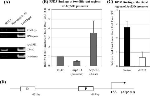
FIG. 4.
dRPD3 binding at the Arp53D promoter region. (A) ChIP assay performed with anti-RPD3 or nonspecific antibodies (Ab). Coprecipitated DNA was analyzed for the presence of promoter sequences of DNA polymerase α (DNApolα), RP49, or two regions of the Arp53D promoter (distal and proximal regions as determined by the presence of putative E2F binding sites and described for panel D). (B) ChIP results from three independent experiments were determined by quantitative real-time PCR. (C) dRPD3 binding is dE2F2 dependent. ChIP assay was performed with anti-RPD3 antibodies in white (control) or dE2F2-depleted cells. ds, double stranded. (D) Structure of Arp53D promoter region. D, distal region; P, proximal region; TSS, transcription start site. The numbers depict the distance from the TSS.

