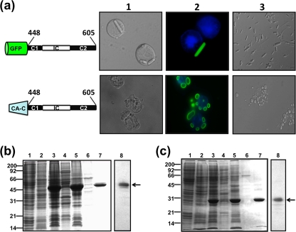
FIG. 7.
Baculovirus expression and purification of μNS-Mi chimeras. (a) Sf9 cells were infected with baculoviruses expressing the chimeras indicated on the left. The cells were fixed 3 days after infection and then either treated with DAPI to stain the nuclei (top row) or stained with DAPI and anti-μNS antibodies and photographed under a bright-field (column 1) or fluorescence (column 2) microscope. Column 3 shows images of the tubular (top) or globular (bottom) inclusions purified from the cytoplasm of infected Sf9 cells. (b) Expression, purification, and immunoblot analysis of tubular inclusions (GFP-C1-IC-C2). Sf9 cells infected with baculovirus expressing GFP-C1-IC-C2 were lysed in hypotonic buffer at 72 h p.i., and the resulting cell extract (lane 3) was fractionated by centrifugation into pellet and supernatant fractions (the supernatant fraction is shown in lane 4). The pellet was washed twice with hypotonic buffer, resuspended in the same volume of hypotonic buffer, and sonicated. The sonicated extract (lane 5) was centrifuged and fractionated into pellet and supernatant fractions (the supernatant fraction is shown in lane 6). The pellet was then washed and centrifuged five times with hypotonic buffer (lane 7). Mock-infected or wild-type-baculovirus-infected Sf9 cell extracts lysed in hypotonic buffer at 72 h p.i. are shown in lanes 1 and 2, respectively. All samples were resolved by 12.5% SDS-PAGE, and the protein bands were visualized by Coomassie blue staining. The position of recombinant GFP-C1-IC-C2 is indicated by an arrow on the right and those of the molecular weight markers on the left. The sample in lane 7 (purified tubular inclusions) was subjected to Western blot analysis with anti-μNS antibodies (lane 8). (c) Expression, purification, and immunoblot analysis of globular inclusions (CA-C-C1-IC-C2). The expression and purification of globular inclusions was performed as for panel b. The position of recombinant CA-C-C1-IC-C2 is indicated by an arrow on the right and those of the protein markers on the left. The sample in lane 7 (purified globular inclusions) was subjected to Western blot analysis with anti-μNS antibodies (lane 8).

