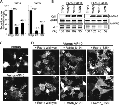
FIG. 2.
Dominant-negative mutants of Rab1b reduce Ebolavirus VLP production. (A) Inhibition of secretory pathways by dominant-negative mutants of Rab1a or Rab1b. Secreted alkaline phosphatase (SEAP) was coexpressed with Rab1a, Rab1b, or their dominant-negative mutants in 293 cells. Twenty-four hours posttransfection, SEAP activities were measured. For cells expressing wild-type Rab1 proteins, the secretion index (i.e., the ratio of SEAP activity detected in the culture supernatant to the cell-associated SEAP activity) was defined as 100%. Experiments were carried out in triplicate. (B) Ebolavirus VP40-induced VLP production is reduced by dominant-negative Rab1b mutants. VP40 was coexpressed in 293T cells in the absence (“Empty”) or presence of FLAG-Rab1a, FLAG-Rab1b, or their dominant-negative mutants (“N124I” or “S22N” and “N121I” or “S22N,” respectively). Twenty-four hours posttransfection, the released VLPs and total cell lysates were analyzed by Western blot analysis with an anti-FLAG antibody or an anti-VP40 antibody. The intensities of the VP40 double bands were quantified, and VLP release efficiencies were calculated based on the ratio of VP40 in VLPs and cell lysates. The values obtained with wild-type Rab1a and Rab1b proteins were set to 100%. The results shown are representative of three independent experiments. (C) Morphological changes in cells expressing Venus-VP40 fusion protein. The reporter protein Venus or Venus-VP40 fusion protein was expressed in 293 cells. Twenty-four hours posttransfection, the cells were imaged by using confocal microscopy. Arrowheads indicate filamentous cell protrusions. (D) Dominant-negative Rab1b mutants reduce the frequency of VP40-induced formation of cell protrusions. Venus-VP40 was coexpressed with wild-type or dominant-negative Rab1 proteins in 293 cells. The cells were imaged 24 h posttransfection by using confocal microscopy.

