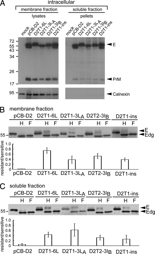
FIG. 8.
Subcellular fractionation experiment of TMD mutants with increased production of VLPs. (A) 293T cells transfected with mock and PrM/E expression constructs were resuspended in modified buffer B and frozen-thawed (27). After clearing the nuclei and debris, the membrane fraction and the pellets derived from the soluble fraction by 20% sucrose cushion ultracentrifugation were subjected to Western blot analysis by using serum from a confirmed DENV2 case (upper) (70) and then reprobed with anti-calnexin MAb (lower). Arrowheads indicate PrM, E, and calnexin. (B) Membrane fraction and (C) pellets of the soluble fraction of each transfectant were treated with endo H (H) or PNGase F (F) and subjected to Western blot analysis by using serum from a confirmed DENV2 case (70). The ratio of the intensity of the endo H-resistant band to that of the endo H-sensitive band is shown below the gels. Arrowheads indicate E or deglycosylated E protein (Edg). Molecular mass markers are shown in kDa. One representative experiment of two is shown.

