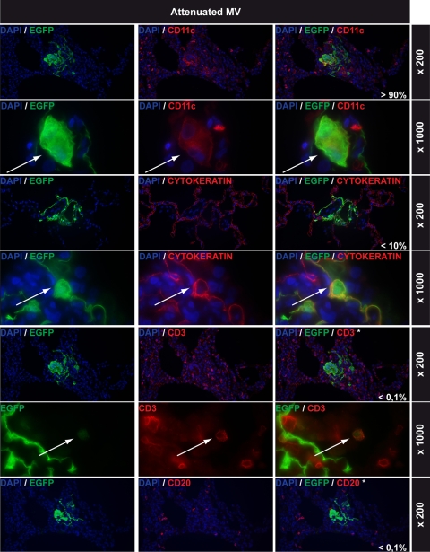
FIG. 5.
Lung infections with attenuated MV: immunohistochemical double stainings. All rows show EGFP staining in green on the left, cell-type-specific staining in red in the middle (CD11c, cytokeratin, CD3, and CD20 for myeloid cells, epithelial cells, T lymphocytes, and B lymphocytes, respectively), and double staining on the right. DAPI counterstaining was used to identify nuclei. Magnifications are indicated on the right of each row. The percentages shown in white in the right-hand panels represent estimations of the relative contributions of these cell types to the total number of MV-infected cells in the lungs. Arrows indicate double-positive cells. Note that EGFP+ cells shown in the CD3 and CD20 double stains are MV-infected CD11c+ cells and that none of the CD3+ or CD20+ cells were EGFP+.

