FIG. 2.
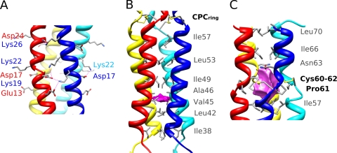
Detailed view of P3 subdomains. (A) Detailed view of the intermolecular salt bridges in the canonical coiled-coil subdomain 1 of P3. Salt bridges are indicated as dotted lines. (B) Lateral view of P3 subdomain 2 revealing the small cavity (magenta) and the hydrophobic layers of the right-handed coiled-coil core, rendered as gray sticks. (C) View of the P3 structure in the region of the 4 cysteine bridges. The CPC ring motif delineates a large cavity (magenta) along the tetramer axis, and the lining residues are shown as sticks.

