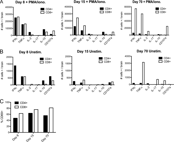
FIG. 6.
CD4+ T cells are the main producers of T-cell-associated IFN-γ within the brains of V3533-infected μMT mice. Female μMT mice (7 to 10 weeks old) were inoculated with 103 PFU of V3533 by injection in the left rear footpad. At the times indicated, mice were perfused with PBS and brain-infiltrating leukocytes were isolated. (A and B) Harvested cells were then pooled and either cultured in the presence of PMA-ionomycin for 6 h with brefeldin A with or without monensin added for the final 4 h (A) or cultured in the presence of brefeldin A with or without monensin with no additional stimulus for 4 h (B). Following treatment, cells were surface stained for CD3α, CD4, and CD8 and then stained for the intracellular presence of multiple cytokines. Each bar represents the number of cells of a given cell type that stained as positive for the indicated cytokine-surface marker per brain (days 8 and 15) or the percentage of pooled cells that stained as positive (day 70). (C) Percentages of T cells positive for CD69 surface expression in the absence of ex vivo stimulation. Data shown were generated in a single experiment but are representative of 2 to 3 independent experiments.

