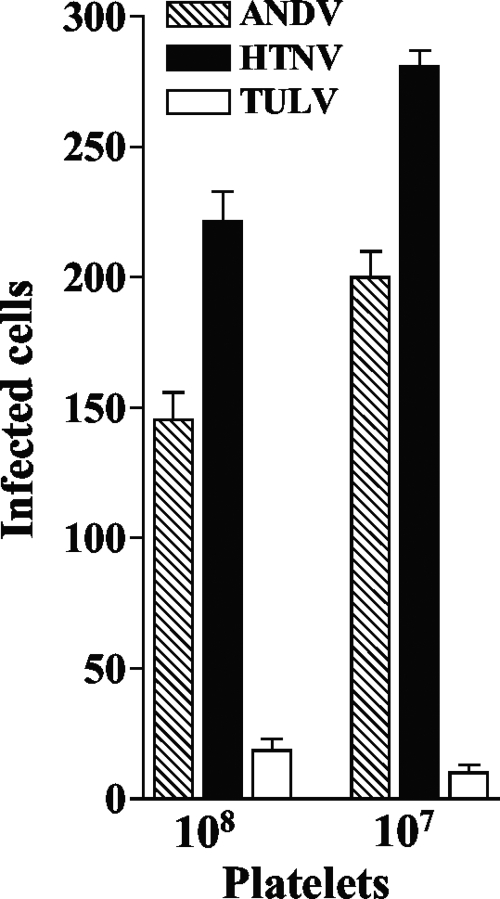
FIG. 1.
Pathogenic hantaviruses bind platelets. Platelets were collected in sodium citrate (final concentration, 0.4%) supplemented with 1 μM prostaglandin E1 (PGE1) (Calbiochem) (27), and platelet-rich plasma (PRP) was prepared by centrifugation for 15 min at 1,800 rpm (700 × g) at 25°C. PRP was supplemented with 10 mM EDTA, and platelets were pelleted by centrifugation (15 min at 1,300 × g at 25°C) and washed with HBMT buffer (HEPES-buffered modified Tyrodes buffer; 10 mM HEPES, pH 7.45, 137 mM NaCl, 2.7 mM KCl, 0.4 mM NaH2PO4, 12 mM NaHCO3, 1 mM MgCl2, 0.1% dextrose, 0.2% bovine serum albumin [BSA] with 1 μM PGE1,10 mM EDTA) (27). Platelets were purified over a Sepharose 2B column equilibrated with HBMT at room temperature and quantitated using a hemacytometer (27). Platelets (107 or 108 per ml) were incubated with 104 FFU of HTNV, ANDV, or TULV for 1 h in an incubator at 37°C and 5% CO2. Platelets were pelleted and washed three times with 10 volumes of HBMT, resuspended in 100 μl HBMT, and then adsorbed onto VeroE6 cells in duplicate wells of a 96-well plate (1 h at 37°C). Monolayers were washed with Dulbecco's modified Eagle's medium supplemented with 2% fetal calf serum, and VeroE6 cells were incubated (37°C, 5% CO2) for 24 h prior to methanol fixation (100%, −20°C) (24). The hantavirus nucleocapsid protein present in infected cells was detected by immunoperoxidase staining as previously described, and the number of N protein-containing cells was quantitated (26). Error bars represent the means ± standard deviations (n = 12) of results from four independent experiments.

