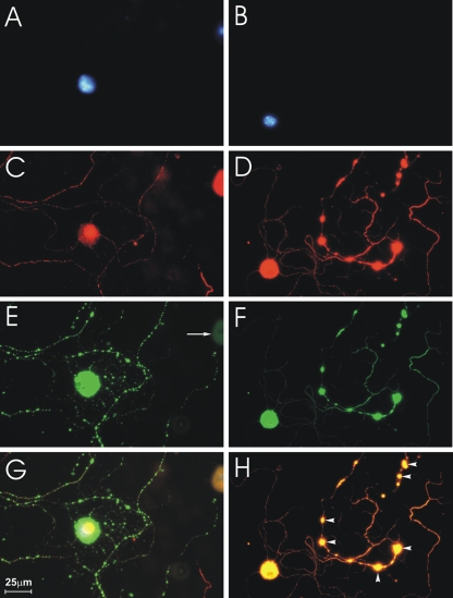
FIG. 1.
CVS infection causes formation of axonal swellings in DRG cultures. Fluorescence microscopy showing CVS-infected DRG neurons at 24 h (A, C, E, and G) and at 72 h (B, D, F, and H) p.i. Staining with DAPI shows neuronal nuclei (A and B). Staining for β-tubulin III shows two neuronal cell bodies at 24 h p.i. (C) and one (large spherical body) at 72 h p.i. (D). (E) There is strong rabies virus antigen staining of one of the two neuronal cell bodies at 24 h p.i., but not of the other, demonstrating that CVS infects only a subpopulation of DRG neurons. Definite axonal swellings are not yet present at 24 h p.i. (C, E, and G), but axonal swellings are well established at 72 h p.i. (D, F, and H, arrowheads). Rabies virus antigen is strongly expressed in the neuronal cell bodies and axons at 24 h and 72 h p.i. (E to H) and also in axonal swellings at 72 h p.i. (F and H).

