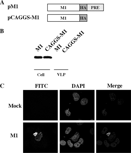
FIG. 1.
M1 alone is not sufficient for extracellular VLP formation. (A) Schematic diagram of M1 expression constructs. An HA epitope tag (YPYDVPDYA) was appended in frame to the C terminus of the M1 protein in pPRE and pCAGGS vectors, producing pM1 and pCAGGS-M1, respectively. (B) VLP production by COS-1 cells transfected with pM1 and pCAGGS-M1 plasmids. VLPs and cell lysates were analyzed by Western blotting with an anti-HA monoclonal antibody. (C) Representative M1 subcellular localization images in transfected COS-1 cells expressing M1 protein. At 48 h posttransfection, COS-1 cells transfected with either pM1 or empty vector (mock) plasmids were fixed, permeabilized, and stained with goat anti-M1 IgG-FITC. Nuclei were stained with DAPI. M1 localization was then examined using confocal microscopy at an ×60 magnification.

