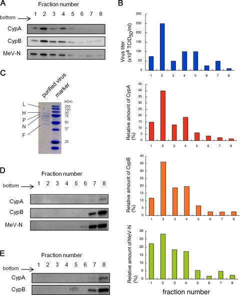
FIG. 3.
Evidence of incorporation of CypA and CypB into MeV particles. Culture medium of MeV-infected B95a cells was ultracentrifuged to pellet down the virus. The resultant pellet was resuspended with TN buffer and placed on a 20% to 60% sucrose layer (the volume of each sucrose layer was 1 ml). After ultracentrifugation, 600-μl fractions were collected sequentially from the bottom of the tube in the case of MeV-HL. (A) CypA, CypB, and MeV-N in each fraction of MeV-HL were detected by Western blotting. (B) The virus titer of each fraction was estimated using B95a cells. The virus titer and relative amounts of CypA, CypB, and MeV-N in each fraction of MeV-HL were graphed. (C) Trichloroacetic acid was added to fraction 2, and the resultant pellet was analyzed by SDS-PAGE and Coomassie brilliant blue staining. MeV-infected or mock-infected B95a cells were lysed with PBS containing 0.5% NP-40, and the cell lysates were placed on a 20% to 60% sucrose layer (the volume of each sucrose layer was 1 ml). After ultracentrifugation, 600-μl fractions were collected sequentially from the bottom of the tube. CypA, CypB, and MeV-N in each fraction of the MeV-infected cell lysate (D) and CypA and CypB in each fraction of the mock-infected cell lysate (E) were detected by Western blotting.

