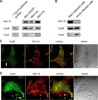
FIG. 4.
Incorporation of CypA or CypB into MeV. (A) MeV-HL, MeV-EGFP, and MeV-luc were purified by sucrose density gradient centrifugation. Virus particles were developed by SDS-PAGE and analyzed by Western blotting with anti-MeV-N, -CypA, and -CypB antibodies. (B) B95a cells were infected with MeV-luc at an MOI of 0.2. At 18 hpi, the cytoplasmic fraction of infected cells was extracted with hypotonic buffer. CypA, CypB, and MeV-N in the cytoplasmic fraction were detected by Western blotting. As a control, mock-infected B95a cells were used. (C and D) Localization of cyclophilins and MeV-N in MeV-luc-infected B95a cells was examined by immunofluorescence staining. CypB (C) and CypA (D) distributions (green; left panels), MeV-N distribution (red; second panels), their overlay (third panels), and phase-contrast images (right panels) are shown in striatal sections. Each panel represents sequential confocal scans of the same field. Arrows in the left panel indicate uninfected cells.

