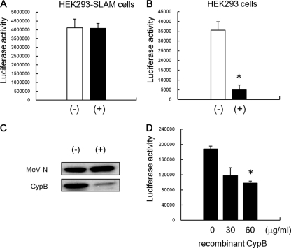
FIG. 5.
Effect of CsA treatment on MeV infectivity to epithelial cells. MeV-luc was propagated with and without 5 μg/ml CsA, and then the culture medium of infected cells was ultracentrifuged to pellet down viruses. The resultant pellets were rinsed and resuspended with fresh medium. The virus propagated without CsA was named MeV-luc (−), and that propagated with CsA was named MeV-luc (+). MeV-luc (−) and MeV-luc (+) were titrated using B95a cells. HEK293-SLAM cells (A) and HEK293 cells (B) were infected with the viruses at an MOI of 1.0. At 24 hpi, the luciferase activity of infected cells was measured. The white and black bars represent the infectivities of MeV-luc (−) and MeV-luc (+), respectively. Luciferase assays were performed in triplicate. Data are means plus standard errors of the means (SEM). *, P = 0.003 (Student's t test). (C) MeV-luc (−) and MeV-luc (+) were further purified by sucrose gradient density centrifugation and were analyzed by Western blotting with anti-MeV-N, -CypA, and -CypB antibodies. (D) HEK293 cells (6 × 10⁴ cells) were preincubated with 0, 30, or 60 μg/ml of recombinant CypB for 30 min at room temperature and then infected with MeV-luc at an MOI of 1.0 in the presence of 0, 30, or 60 μg/ml of recombinant CypB at 37°C for another hour. After incubation, cells were washed three times with medium containing fusion block peptide to stop infection and to remove viruses and recombinant CypB. The luciferase activity of infected cells was measured at 24 hpi. Luciferase assays were performed in triplicate. Data are means plus SEM. *, P = 0.0007 (Student's t test).

