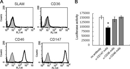
FIG. 6.
Contribution of CD147 to MeV infection of epithelial cells. (A) HEK293 cells (1 × 10⁶ cells) were incubated with 1 μg of anti-CD147, -CD36, -CD46, or -SLAM mouse monoclonal antibody for 1 hour. After being washed, cells were incubated with 0.5 μl of Alexa Fluor 468-conjugated goat anti-mouse IgG polyclonal antibody for 1 hour. After being washed, cells were analyzed by flow cytometry. (B) HEK293 cells (6 × 10⁴ cells) were preincubated with 50 μg/ml of anti-CD147, -CD36, or -CD46 antibody for 30 min at room temperature and then infected with MeV-luc at an MOI of 1.0 in the presence of 50 μg/ml of each antibody at 37°C for another hour. After incubation, cells were washed three times with medium containing fusion block peptide to stop infection and to remove viruses and antibodies. The luciferase activity of infected cells was measured at 24 hpi. Luciferase assays were performed in triplicate. Data are means plus SEM. *, P = 0.020 (Student's t test).

