FIG. 2.
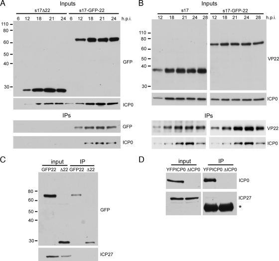
VP22 and ICP0 form a complex in infected cells. (A) Vero cells infected with s17 expressing GFP in place of VP22 (s17Δ22) or expressing GFP-tagged VP22 (s17-GFP-22) at a multiplicity of infection of 1 were harvested at times ranging between 6 and 24 h after infection, and immunoprecipitations carried out with a VP22 polyclonal antibody. The original input samples and resulting complexes were analyzed by Western blotting for GFP or ICP0. The sizes of the molecular weight markers (in thousands) are indicated on the left. (B) As described in the legend for panel A, but using s17 and s17-GFP-22 viruses. (C) Vero cells infected with the same viruses as those shown in panel A were harvested 20 h after infection and immunoprecipitated with a polyclonal anti-GFP antibody. The resulting complexes were analyzed by Western blotting for GFP and ICP27. (D) Vero cells infected with either s17 expressing YFP-tagged ICP0 or the ICP0 deletion virus dl1403 (ΔICP0) were treated as described in the legend for panel C and analyzed by Western blotting for ICP0 and ICP27. Asterisk denotes polyclonal heavy chain.

