FIG. 3.
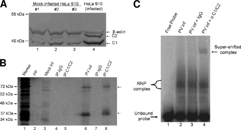
The cellular protein hnRNP C binds the 5′ end of poliovirus negative-strand RNA. (A) Increased concentrations of hnRNP C proteins in HeLa S10 cytoplasmic extracts generated from poliovirus-infected cells. Cytoplasmic extracts of HeLa cells were prepared from either mock-infected cells (three separate extracts; lanes 1 to 3) or poliovirus-infected cells (lane 4) at 5 h postinfection. Equivalent amounts of total protein (10 μg) from each extract were resolved on a 12.5% polyacrylamide-SDS-containing gel. Western blot analysis was carried out using mouse monoclonal anti-C1/C2 as the primary antibody (Abcam) and an alkaline phosphatase-conjugated anti-mouse IgG (Zymed) as a secondary antibody. Molecular masses are indicated on the left. As a loading control, the blots were also probed with mouse monoclonal anti-β-actin antibody (Abcam). (B) Immunoprecipitation of UV cross-linked complexes confirms binding of hnRNP C to the 5′(−) RNA probe. UV cross-linking assays were carried out as described in Materials and Methods with the [32P]UTP-radiolabeled 5′(−) RNA probe and cytoplasmic extracts generated from mock-infected (Mock inf) (lanes 3 to 5) or poliovirus-infected (PV inf) (lanes 6 to 8) HeLa cells at 5 h postinfection. [32P]UTP-radiolabeled UV cross-linked samples from mock- or poliovirus-infected HeLa cells were immunoprecipitated by the inclusion of normal IgG (lanes 4 and 7) or hnRNP C1/C2 antibody (lanes 5 and 8) in reaction mixtures following RNase digestion. The immunoprecipitated complexes were resolved on a 12.5% polyacrylamide-SDS-containing gel. Analysis was carried out using a phosphorimager. Lane M, marker proteins ([35S]methionine-labeled in vitro translation of poliovirus virion RNA). FP, free probe in the absence of any extract. (C) Electrophoretic mobility shift assays using extracts generated from poliovirus-infected cells in the presence or absence of antibody to analyze the contents of RNP complexes. Cytoplasmic extracts (1 μg) from poliovirus-infected HeLa cells were incubated with radiolabeled poliovirus 5′(−) RNA probe. The reaction mixtures in lanes 3 and 4 contained 0.1 μg of either IgG or C1/C2 monoclonal antibody, respectively. The unbound probe is indicated at the bottom left, and the appearance of a supershifted complex in lane 4 is indicated by an arrow at the top right.

