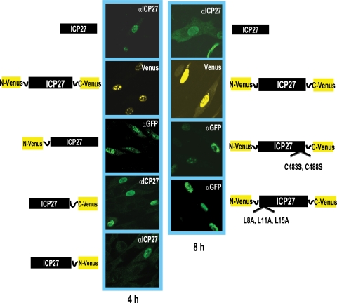
FIG. 2.
Immunofluorescence analysis indicates that the ICP27-Venus fusion constructs are expressed and localized to the nucleus. RSF cells were transfected with plasmid DNA from a wild-type ICP27-containing plasmid and from the ICP27-Venus fusion constructs indicated. Eighteen hours after transfection, cells were infected with 27-LacZ at an MOI of 10 for 4 and 8 h as indicated. Wild-type ICP27 was stained with anti-ICP27 antibody P1119, as was ICP27-C-Venus and ICP27-N-Venus. N-Venus-ICP27, N-Venus/ICP27 (C483S, C488S)/C-Venus, and N-Venus/ICP27 (L8A, L11A, L15A)/C-Venus were stained with an anti-GFP antibody that recognizes an epitope also present in Venus. The P1119 anti-ICP27 antibody recognizes an epitope at the N terminus of ICP27 (23) that is masked by the N-Venus fusion protein. Venus yellow fluorescence was visualized directly for N-Venus/ICP27/C-Venus.

