FIG. 7.
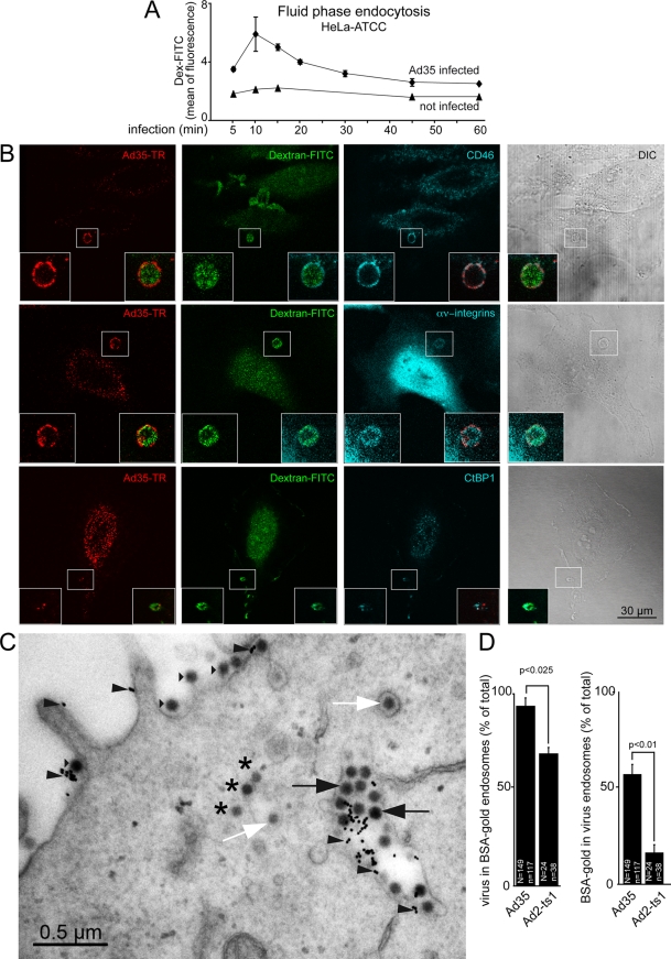
Ad35-TR induces fluid phase uptake and colocalizes with CD46, integrins, and CtBP1 in dextran-filled macropinosomes. (A) Ad35 (2 mg/ml) was cold bound to HeLa-ATCC cells for 1 h. Cells were washed, pulsed with dextran-FITC (0.5 mg/ml) in warm medium (37°C) containing BSA 5 min before the indicated time points, and prepared for flow cytometric analysis. (B) Cells were cold bound with Ad35-TR (1 μg/ml) for 1 h, washed, and incubated at 37°C in the presence of dextran-FITC (0.5 mg/ml) for 10 min, fixed, stained for the indicated antigens, and analyzed by confocal laser scanning microscopy with corresponding differential interference contrast (DIC) images. Fluorescence images represent single sections, and insets are magnifications of the white boxed areas. (C) Electron micrograph of an Ad35-infected HeLa-ATCC cell (cold synchronized infection at a MOI of 5,000), pulsed in the presence of BSA-nanogold for 10 min. Arrowheads indicate BSA-gold particles, black arrows show Ad35 in BSA-gold-positive endosomes, white arrows show virus particles in endosomes without BSA-gold, small triangles indicate viruses at the plasma membrane, and stars depict Ad35 particles in the cytosol. (D) Quantification of panel C, including HeLa-ATCC cells inoculated with Ad2-ts1, which is defective in endosomal escape. “N” indicates the number of virus particles, and “n” indicates the number of BSA-gold particles.

