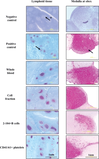
FIG. 1.
Terminal lymphoid and brain (medulla at obex) IHC analysis results of naïve deer cohorts inoculated with CWD+ blood components. PrPCWD demonstrated by IHC analysis in tonsil, brain (medulla oblongata at obex), and retropharyngeal lymph node tissues of deer receiving whole blood, cell fraction, B cells, or CD41/61+ cells from CWD-infected donors. Arrows indicate PrPCWD staining (red) within the brain and lymphoid follicles. The arrow with the asterisk indicates a lymphoid follicle negative for PrPCWD.

