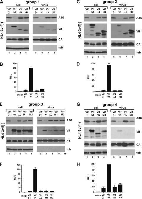
FIG. 2.
Vif mutants are nonfunctional. (A) 293T cells were transfected with 3 μg of pNL4-3vif(−) virus and 0.5 μg of pcDNA-A3G, together with 2.5 μg each of empty vector (lanes 1 and 5), pcDNA-hVif wt (lanes 2 and 6), Vif-ΔK (lanes 3 and 7), or Vif-ΔD (lanes 4 and 8). Cells and virus-containing supernatants were harvested 24 h posttransfection. Whole-cell lysates and virus-containing supernatants were prepared as described in Materials and Methods. Samples were analyzed by immunoblotting them using antibodies to A3G and Vif. The Vif blot was subsequently reprobed with an HIV-positive patient serum to detect viral capsid protein (CA), and the A3G blot was reprobed with a tubulin-specific monoclonal antibody (tub). (B) The infectivities of the viruses shown in panel A were determined in a single-cycle infectivity assay by infection of LuSIV indicator cells (52). Virus-induced activation of luciferase, as measured in relative light units (RLU), was quantified 24 h later in a standard luciferase assay as described in Materials and Methods. The relative infectivity of viruses produced in the presence of Vif wt was defined as 100%. Uninfected LuSIV cells were included as a negative control (mock). The error bars reflect standard deviations calculated from three independent infections. (C and D) 293T cells were transfected as for panel A, except that Vif-ΔJ and Vif-ΔH mutants were analyzed. Analysis of viral infectivity was done as for panel B. (E and F) 293T cells were transfected as for panel A, except that Vif-ΔC, Vif-M1, and Vif-M2 mutants were analyzed. The infectivities of the resulting viruses were determined as for panel B. (G and H) 293T cells were transfected as for panel A, except that Vif-ΔN and Vif-M3 mutants were analyzed. Analysis of viral infectivity was done as for panel B.

