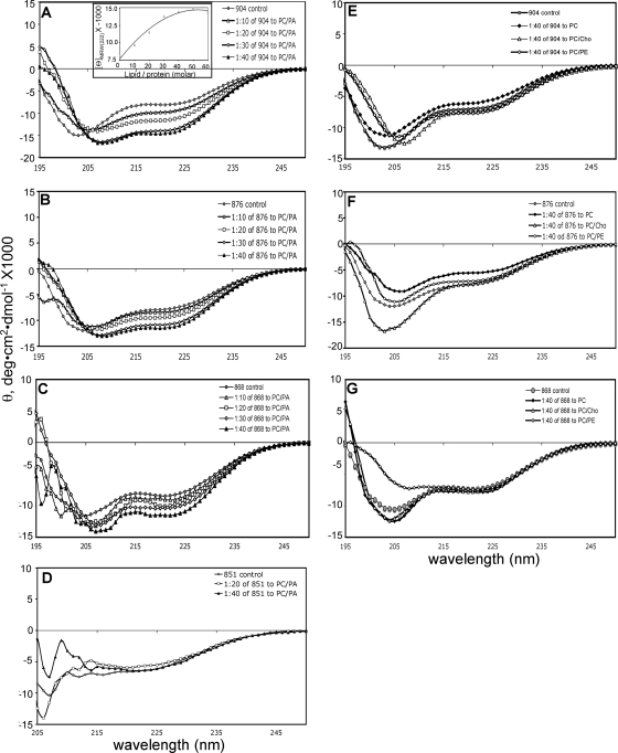
FIG. 4.
Far-UV CD spectra of the full-length and the truncated cytoplasmic domains of HSV-1 gB in the presence of lipid SUVs. (A to D) Spectra recorded in the presence of increasing molar ratios of PC/PA SUVs to protein. (A) gB(801-904); (B) gB(801-876); (C) gB(801-868); (D) gB(801-851). (E to G) Spectra were recorded in the presence of a 40:1 molar ratio of PC, PC/PE, or PC/Chol SUVs to protein. (E) gB(801-904); (F) gB(801-876); (G) gB(801-868). Control spectra in each panel are of protein in buffer alone. The inset in panel A shows θ at 222 nm plotted against increasing molar ratios of PC/PA SUVs to protein.

