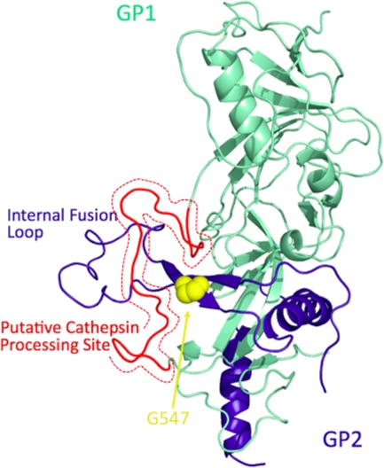
FIG. 5.
Three-dimensional structure of the Angola GP monomer. The crystal structure of EBOV GP (PDB code 3CSY) was used as a template for homology modeling. GP1 (lime green) and GP2 (dark blue) are shown as ribbon models. Glycine at position 547 (G547, yellow) is shown as a space-filling model. The putative cathepsin cleavage site (amino acid residues 174 to 197 of Angola GP, corresponding to amino acid residues 190 to 213 of Zaire EBOV GP) is colored in red. This figure was prepared using PyMOL (DeLano Scientific LLC).

