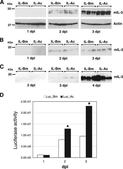
FIG. 6.
Expression of mIL-3 and luciferase using a novel BmNPV vector with AcMNPV fp25K. (A) Expression of mIL-3 in BmN cells. BmN cells were infected with IL-Bm and IL-Ac and harvested at 1 to 3 dpi in cell lysis buffer. The lysates were then analyzed by Western blotting with anti-FLAG antibody. Western blotting using anti-actin was also performed. The molecular masses of protein standards are indicated to the left. (B) Secretion of mIL-3 in BmN cells. BmN cells were infected with IL-Bm and IL-Ac, and the culture medium was harvested at 1 to 3 dpi. Western blotting of the medium samples was performed with anti-FLAG antibody. The molecular masses of protein standards are indicated to the left. (C) Secretion of mIL-3 into the larval hemolymph. B. mori larvae were injected with BVs from IL-Bm and IL-Ac, and hemolymph samples were collected at 2 to 4 dpi. Western blot analysis of the hemolymph samples was performed with anti-FLAG antibody. (D) Expression of luciferase in BmN cells. BmN cells were infected with Luc-Bm and Luc-Ac and harvested at 1 to 3 dpi in cell lysis buffer. The lysates were then subjected to a luciferase assay. Data shown are means ± SD (n = 3). *, P < 0.05 by Student's t test.

