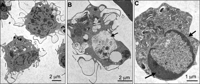
FIG. 3.
Electron micrograph of peritoneal macrophage-like cells from FV3-infected Xenopus adults. PLs were isolated from frogs at 2 days postinfection with 5 × 10⁶ PFU of FV3, processed, and visualized under a Hitachi 7650 TEM. (A) Two mononucleated macrophage-like cells with multiple pseudopods. (B) A macrophage-like cell with a large phagocytic vacuole (arrow). (C) Apoptotic cells showing heterochromatic condensation (arrows).

