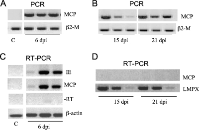
FIG. 7.
In vivo FV3 infection and transcription in PLs. PCR analysis (35 cycles) was performed on total DNA (0.5 to 1 μg) from PLs isolated from three animals infected for 6 days (A) or for 15 and 21 days (B) with 1 × 10⁶ PFU of FV3 and using primers specific for the FV3 MCP gene and β2-microglobulin (β2-M) as positive controls. RT-PCR analysis was performed on total mRNA (500 ng) isolated from uninfected or infected PLs at 6 days (C) or at 15 and 21 days (D) postinfection using primers for the MCP and IE genes of FV3 and β-actin or LMPX (large multifunctional protease X) as controls for 30 cycles. Data shown are representative of two experiments. −RT, controls lacking reverse transcriptase.

