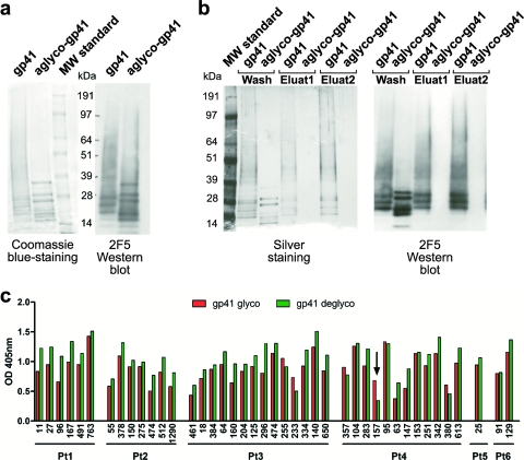
FIG. 6.
Rare glyco-dependent binding by anti-gp41 antibodies. (a) Coomassie blue-stained polyacrylamide gel and Western blot analysis with MAb 2F5 of control and deglycosylated gp41. As specified by the manufacturer (Acris), the protein shows three major immunospecific bands between 20 and 30 kDa, minor bands between 20 and 30 kDa, bands at 14 and 7 kDa, and an aggregation smear at 35 kDa and above. The aggregation smear condenses in major specific bands after deglycosylation. (b) Deglycosylation was further confirmed by Lens culinaris lectin precipitation, followed by elution with 0.5 M methyl α-d-mannopyranoside of glycosylated gp41 but not of the deglycosylated form. (c) Graph summarizes differences in binding of anti-gp41 antibodies to intact (red bars) and deglycosylated (green bars) gp41 as measured by ELISA (OD at 405 nm). A significant decrease in binding is indicated by a black arrow.

