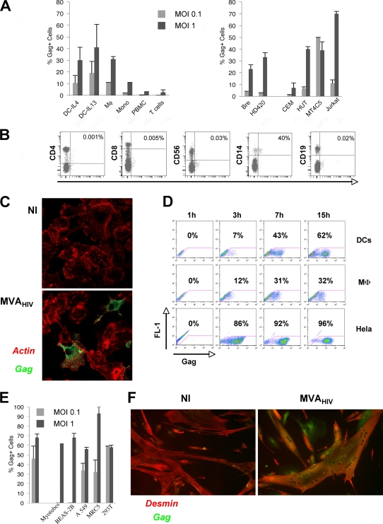
FIG. 1.
MVA-HIV preferentially infects APCs and vaccination target cells. (A) Gag expression in primary cells (left) or in cell lines (right) after MVA-HIV infection at the indicated MOI. At 15 h p.i., cells were stained for intracellular HIV Gag and analyzed by flow cytometry. DCs were differentiated in the presence of IL-4 or IL-13. The data represent the means ± standard deviations (SD) for at least three independent experiments. (B) PBMCs were infected with MVA-HIV (MOI = 1). At 15 h p.i., cells were stained for cell surface markers and intracellular HIV Gag and analyzed by flow cytometry. Uninfected PBMCs and isotype-matched Abs were used as negative controls (not shown). T cells, CD8+ and CD4+ cells; NK cells, CD56+ cells; monocytes, CD14+ cells; B cells, CD19+ cells. The percentages of Gag+ cells in the corresponding cell subsets are shown. The data are representative of three independent experiments. (C) DCs were infected with MVA-HIV (MOI = 0.1), and HIV Gag expression was further analyzed by immunofluorescence and confocal microscopy. Green, HIV Gag staining; red, actin-phalloidin-PE. NI, not infected. (D) Kinetics of HIV Gag expression in DCs, macrophages (MD-M), and HeLa cells analyzed as described for panel A. (E) HIV Gag expression in primary myotubes and epithelial cells 15 h after MVA-HIV infection (MOI = 1), assessed using flow cytometry and immunofluorescence for myotubes. The data represent the means ± SD for at least three independent experiments. (F) Myotubes were infected with MVA-HIV (MOI = 1), and HIV Gag expression was analyzed by immunofluorescence and confocal microscopy. Green, HIV Gag staining; red, anti-desmin MAb to identify myotubes. NI, not infected.

