FIG. 3.
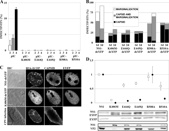
Functions of mutated NS1 constructs. (A) Secondary infections with concentrated medium collected at 2, 3, and 4 days p.t. are shown with white, gray, and black bars, respectively. Infectivity is the percentage of NS1-positive nuclei in the sample (n > 5,100). Error bars indicate standard deviations. (B) Percentages of transfected cells with signs of infection (n = 60). Chromatin marginalization is indicated in white, nuclear capsid antibody staining in black, and cells with both markers in gray. (C) NS1-deYFP-expressing infected cell with marginalized chromatin and nuclear capsid accumulation, K406M-deYFP-expressing infected cell with nuclear capsid accumulation but normal chromatin distribution, and CPV-infected cell, fixed at 48 h p.i., with marginalized chromatin and cytoplasmic capsid localization. Bars, 5 μm. DIC, differential interference contrast. (D) P38 transactivation activities of mutated NS1 proteins. Average transactivation values from EYFP (white boxes) and pIC (black circles) experiments are shown (n = 5). Transactivation values are average values for the product under the P38 promoter (EYFP or VP2) divided by the value for the corresponding NS1 protein (NS1-EYFP or NS1) and normalized to the value for nonmutated NS1. Proteins were detected in Western blots with anti-GFP (NS1-EYFP and EYFP), anti-NS1 (NS1), or anti-capsid (VP2) antibody. Error bars indicate standard errors of the means.

