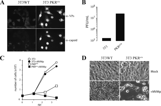
FIG. 1.
Analysis of MVMp replication in NIH 3T3 and 3T3 PKRo/o cells. (A) IF of MVMp-infected cells probed at 24 hpi with specific antibodies for detection of total virus structural proteins (α-VPs) or the protein subunits assembled in capsids (α-capsids). (B) MVMp yield in wild-type 3T3 and 3T3 PKRo/o cells. Supernatants of infected cultures (MOI, 0.1) were sampled, and extracellular viral yield was quantified by a plaque assay. A representative result obtained at 48 hpi is shown. (C) Effect of MVMp infection (MOI, 5) on NIH 3T3 and 3T3 PKRo/o cell growth rates. The number of viable cells measured by trypan blue exclusion at the indicated days postinfection (dpi) is shown. (D) Micrographs showing the cytopathic effect provoked by MVMp on the cultures outlined in panel C at 3 dpi.

