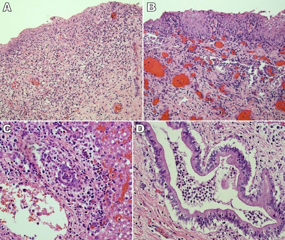
FIG. 2.
Photomicrographs of selected tissue biopsy specimens that were taken at autopsy. (A) Hematoxylin and eosin (H&E) staining (×200 magnification) of a section of the vagina illustrated focal ulceration and granulation tissue with mixed acute and chronic inflammation. There was also focal necrosis of the vaginal tissue. In addition, there were focal collections of neutrophils in blood vessels as well as vascular congestion. Finally, increased intraepithelial lymphocytes, edema, and occasional necrotic keratinocytes were observed. These findings are consistent with those from vaginal tissue taken from a fatal case of tampon-related toxic shock syndrome (1). (B) H&E staining (×200 magnification) of a section of the cervix illustrated mixed acute and chronic ectocervical inflammation. (C) H&E staining (×400 magnification) of a section of liver tissue showed mixed acute and chronic periportal inflammation and acute inflammation in bile ducts. (D) H&E staining (×400 magnification). Acute cholangitis was observed in sections of the bile ducts. These findings are consistent with the pattern of hepatic injury and secondary cholangitis associated with toxic shock syndrome (6).

