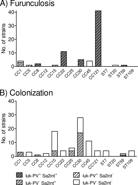
FIG. 3.
Distribution of luk-PV genes and PVL-encoding phage Sa2int within the spa-defined CCs among furunculosis strains (A) and colonizing strains (B). luk-PV-positive (luk-PV+) phages characterized the furunculosis strains, whereas their luk-PV-negative (luk-PV−) counterparts were frequent among the colonizing strains. The total number of strains per CC is represented by the overall height of the bar, whereas the numbers of strains positive for luk-PV and phage Sa2int (Sa2int+) are represented by stripes and gray shading, respectively. Sa2int−, phage Sa2int negative.

