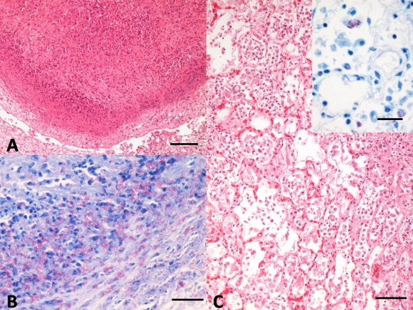
FIG. 2.
(A) Alpaca 1. Nodular granulomatous lesion composed of a central core of necrosis, surrounded by degenerated neutrophils and cell debris, epithelioid macrophages, and scattered lymphocytes and plasma cells. Note the manifest capsule of connective tissue delimiting the lesion (as shown by hematoxylin and eosin [H&E] staining). Bar = 100 μm. (B) Alpaca 1. Abundant acid-fast bacteria in the periphery of the granulomatous lesion. Ziehl-Neelsen staining. Bar = 20 μm. (C) Alpaca 3. Marked proliferation of histiocytes, with diffuse intraalveolar infiltrate of foamy macrophages and epithelioid cells. H-E. Bar = 50 μm. The inset image shows random foamy macrophages, laden with numerous acid-fast bacteria within their cytoplasm. Ziehl-Neelsen staining. Bar = 20 μm.

