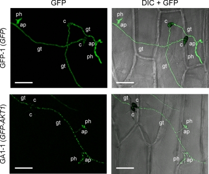
Fig. 4.
Intracellular localization of GFP-tagged Akt1 in infection-related structures. A sample of a conidial suspension was dropped onto onion epidermis and incubated for 24 h. GFP-1, pYTGFPc transformant; GA1-1, pGFP-AKT1 transformant; GFP, GFP fluorescence images; DIC + GFP, DIC images merged with GFP fluorescence images. c, conidium; gt, germ tube; ap, appressorium; ph, penetration hypha. Bars = 50 μm.

