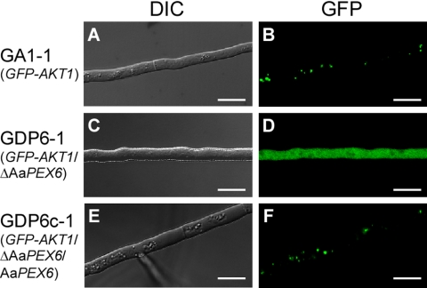
Fig. 6.
Intracellular localization of the GFP-Akt1 fusion protein in hyphal cells of a ΔAaPEX6 mutant strain. Strains were grown on PDA for 3 days. GA1-1, GFP-Akt1-expressing strain (A and B); GDP6-1, ΔAaPEX6 mutant strain made from GA1-1 (C and D); GDP6c-1, AaPEX6-complemented strain made from GDP6-1 (E and F); DIC, DIC images; GFP, GFP fluorescence images. Bars = 10 μm.

