Fig. 2.
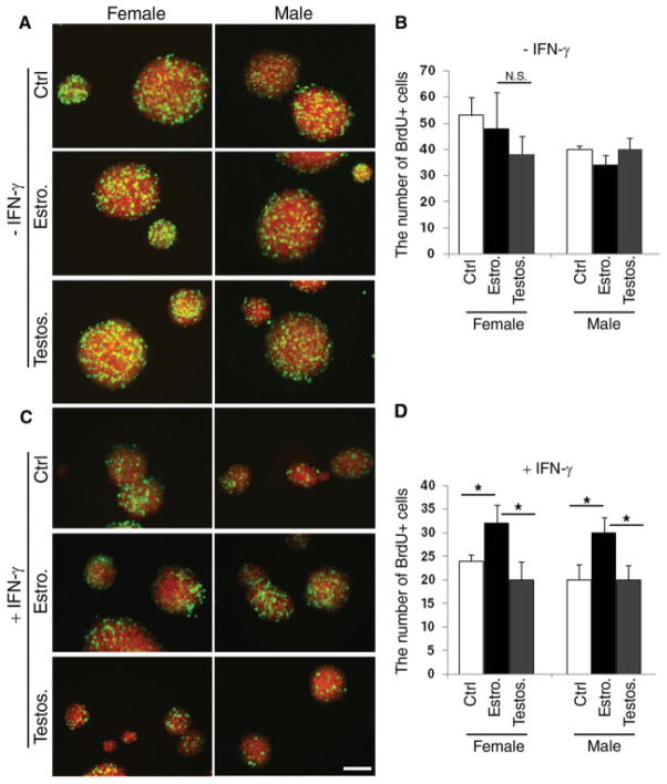
The proliferative response of SVZ cells to hormonal treatment is enhanced by the presence of cytotoxic cytokines. A: Confocal image of secondary neurospheres generated by mechanically dissociating primary neurospheres and growing the same numbers of cells in medium supplemented with EGF alone (ctrl) or with 100 nM estrogen (Estro) or testosterone (Testos) for 7 days. The cells in S-phase were labeled with a 6-hr pulse of BrdU and identified by immunocytochemistry using specific antibodies for BrdU (green) and Sox2 (red). B: Bar graphs represent the results of the quantification of BrdU+ cells in secondary neurospheres cultured as described in A. Bar shows average of three experiments ± SD. C: Confocal image of secondary neurospheres generated by mechanically dissociating primary neurospheres and growing the same numbers of cells in medium supplemented with EGF + 10 ng/ml IFN-γ alone (ctrl) or with 100 nM estrogen (Estro) or testosterone (Testos) for 7 days. Note the dramatic effect of IFN treatment on the size of the male-derived neurospheres. D: Bar graphs represent the results of the quantification of BrdU+ cells in secondary neurospheres cultured as described in C. Bar shows average of three experiments ± SD. *P < 0.005. Scale bar = 100 μm.

