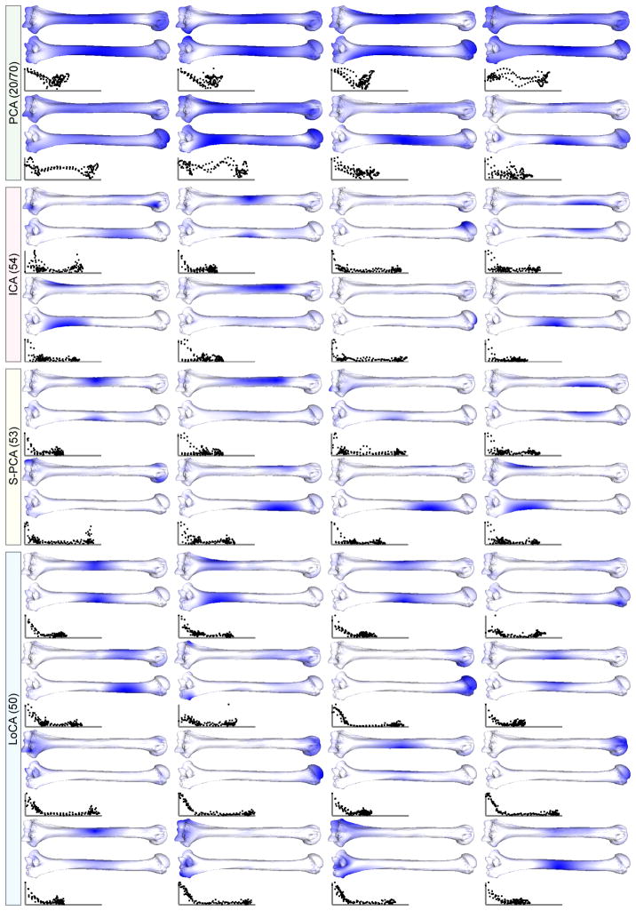
Fig. 5.
Front and back views of displacements on primate humeri. Displacements of the landmark points are computed using different methods, and then interpolated onto the mesh using a thin-plate-spline. Darker locations indicate greater displacement magnitudes. LoCA components describe the articular surfaces at the two extremities as well as deformations of the shaft.

