
Figure 2. Failure of TH17 generation from naive cells of patients with HIES.
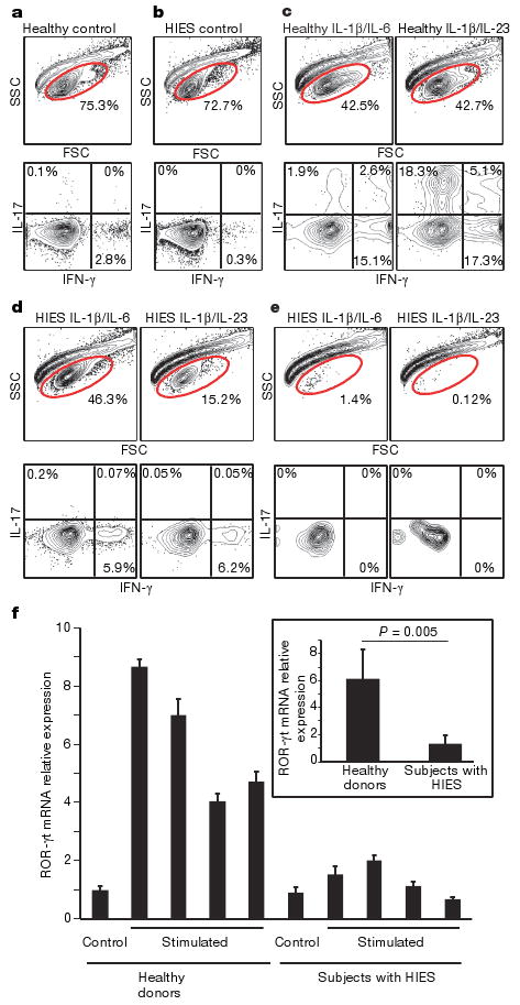
A total of 50,000 naive T cells from healthy controls and patients with HIES were stimulated with anti-CD2/CD3/CD28 microbeads and the indicated cytokines as described in Methods. a–e, Dot plots of intracellular cytokine staining from healthy control (a) and HIES (b) cells stimulated without any cytokines, and from control subjects (c) and subjects with HIES (d, e) stimulated in the presence of IL-6 and IL-1β, or with IL-23 and IL-1β. e, Representative forward and side scatter plots of HIES cells after stimulation for 12 days show the poor recovery after stimulation with either IL-6 and IL-1β, or IL-23 and IL-1β. d, The one subject sample that had good cellular recovery. f, Naive CD4 T cells from four healthy donors and four subjects with HIES were freshly lysed or stimulated with anti-CD3 and anti-CD28 in the presence of IL-23 for 48h. ROR-γt mRNA expression was detected by quantitative RT–PCR. Values shown are relative expression levels of triplicate samples (means and s.d.). Inset: average expression levels for the four healthy donors compared with those for the four subjects with HIES. Statistical significance was determined with a two-tailed unpaired Student t-test.