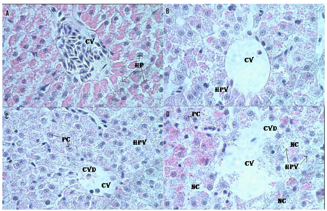
Fig. 5.
Histopathological evaluation of Carassius auratus liver exposed to Cr (VI) for 96 h. A= Control (CV= Central vein, HP=Hepatocytes); B= LC12.5 exposed liver (CV= Central vein, HPV= Hepato cellular vacuolation); C= LC25 exposed liver (CV=Central vein, CVD= Central vein damage, HPV= Hepato cellular vacuolation and PC=Pycknotic); and D= LC50 exposed liver (CV=Central vein, CVD= Central vein damage, NC= Necrosis and PC=Pycknotic). H & E Staining 1000 X.

