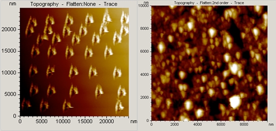
Figure 1.
Atomic force microscopy topographic images. (Left) Nanoindented arrays (25 × 25 μm2) of a clean gold (111) surface. Indents decorate the monatomic gold substrate with “cavities.” (Right) Topographic image (10 × 10 μm2) for the p(HEMA)-PEDOTPEGMA composite hydrogel prepared by electrochemical deposition at +0.850 volt for 100 seconds.

