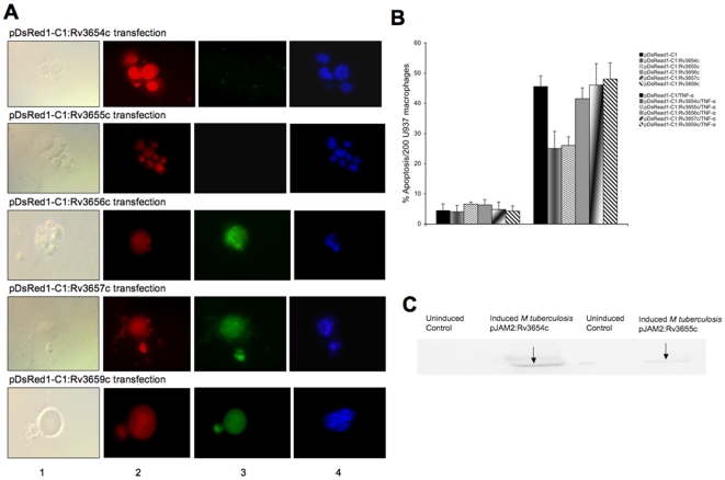
Figure 3. Macrophage transfection affects apoptosis.
A CaspaTag™ In Situ analysis of U937 macrophages for caspase-8 activation and nuclear changes during transfection with M. tuberculosis proteins. While transfection with M. tuberculosis Rv3654c and Rv3655c genes during TNF-α treatment of macrophages suppressed caspase-8 activation and nuclear fragmentation, M. tuberculosis Rv3656c-, Rv3657c- and Rv3659c- transfected and TNF-α-treated macrophages showed caspase-8 activation and nuclear morphological changes typical of apoptosis. Colors in the columns: (1) Phase contrast, (2) Red – transfection, (3) Green – caspase-8 activation and (4) Blue – nuclear staining with Hoechst stain. Bar, 10 µm. B Effect of M. tuberculosis-secreted proteins on caspase-8 activation during macrophage apoptosis. U937 cells were transfected with Rv3654c–Rv3659c genes for 8 h and then treated with human recombinant TNF-α protein (0.5 µM). Active caspase-8 was quantified using In Situ Assay kit (Chemicon). C Analysis of secreted proteins in the cytoplasm of infected macrophages. U937 cells were infected with M. tuberculosis containing pJAM2:Rv3654c or pJAM2:Rv3655c over-expressed vectors at an MOI 1∶10. Concentrated bacterial and cell lysate-free supernatants were subjected to Western blotting using His-tag antibody.

