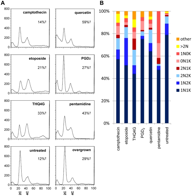
Figure 3. DNA content and genome segregation following drug treatment.
A) DNA content following 24 hour treatment revealed by PI staining of RNAse treated BF. The large peak at left seen in some samples represents cells with degraded DNA. 2C indicates diploid (G1) DNA content, 4C indicates G2/M DNA content. Note the appearance of cells with sub-G1 DNA (peaks at far left) following some treatments, as well as cells with higher order DNA content. In each case, the total number of live cells was less than 50% of the untreated controls. The percentage of dead cells (%†) is indicated on each graph. B) Duplication and segregation of the nucleus and kinetoplast following drug treatment. The same populations of cells analyzed in Figure 3A were subjected to microscopic analysis, enumerating the number of nuclei and kinetoplasts per cell as revealed by DAPI staining. Forms seen during normal cell cycle progression are indicated by blue shades, while red to yellow shades indicate abnormal, dead end forms. N, nucleus, K, kinetoplast.

