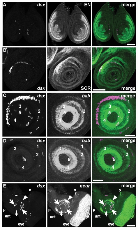
Figure 4. dsxGAL4 is expressed in imaginal discs.
Fluorescence of indicated reporters in imaginal discs of third instar female larvae is shown. (A) dsxGAL4 driving expression of UAS-Stinger nuclear GFP reporter (magenta) is compared to the posterior compartment of the disc marked by EN (green) in this pair of foreleg discs from an early wandering third instar larva. At this stage, few cells express dsxGAL4, but the majority of these cells form a broken crescent within the anterior compartment. Scale bar, 100 µm. (B) Expression of UAS-Stinger nuclear GFP reporter (magenta) in the foreleg disc is compared to the position of tarsal and tibial primordia that express high levels of SCR (green). In this disc from a more mature wandering third instar larva than shown in (A) there are more expressing cells in the dsxGAL4 crescent, and the crescent overlaps with the SCR domain. Scale bar, 100 µm. (C) Expression of UAS-RedStinger nuclear DsRed reporter (magenta) is compared to the position of tarsal segment boundaries defined by concentric expression of bab-lacZ (green). In this foreleg disc from a third instar larva shortly before purarium formation, there is a greater number of cells expressing dsxGAL4 in the dsxGAL4 crescent, and the crescent overlaps with the bab-lacZ domain corresponding to the T1 tarsal segment. Scale bar, 50 µm. (D) Expression of UAS-RedStinger nuclear DsRed reporter (magenta) in a leg disc corresponding to the second or third leg is compared to the position of bab-lacZ (green). In contrast to (A–C), there are only a few cells expressing dsxGAL4 scattered across the disc, and there is no crescent. Scale bar, 50 µm. (E) Expression of UAS-RedStinger nuclear DsRed reporter (magenta) is compared to the position of cells expressing the proneural marker neur-lacZ (green) in the eye-antennal disc from a mature wandering third instar larva. Many cells express dsxGAL4 in the antennal (ant) portion of the disc, while only a few cells express dsxGAL4 in the eye portion. Primordia for the second (arrowhead) and third (barbed arrows) antennal segments are indicated, as are primordia for the arista (arrow), palpus (p), and ocellus (o). Scale bar, 50 µm.

