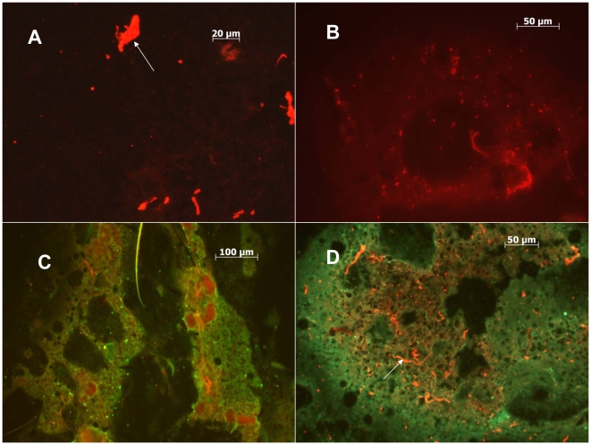
Figure 8. Localization of CxFV Izabal and WNV to head tissues in co-infected mosquitoes.
A) Uninfected head tissues of Cx. quinquefasciatus stained with AlexaFluor 594 (red). White arrow depicts non-specific staining of debris. B) Head tissues of CxFV Izabal-infected Cx. quinquefasciatus, harvested 7 DPI. CxFV Izabal stained with AlexaFluor 594. C) Head tissues of WNV-infected Cx. quinquefasciatus, harvested 9 DPI. WNV stained with AlexaFluor 488 (green). D) Co-infected head tissues of Cx. quinquefasciatus. Mosquito inoculated simultaneously with CxFV Izabal and WNV, harvested 9 DPI. CxFV Izabal stained with AlexaFluor 594 (red) and WNV with Alexa 488 (green). White arrow denotes non-specific staining of debris.

