Fig. 2.
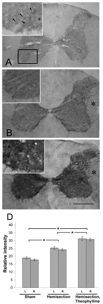
Cytochrome c oxidase activity is increased after spinal cord injury. Photomicrographs depicting the CcO histochemical activity in controlnoninjured, (A), C2HS (B), or C2HST rats (C,). Note that the intensity of the COX reactivity gradually increases from A to C. (*) = contralateral to the injury. In higher power photomicrographs (insets) neuronal-like profiles are occasionally observed (arrowheads in A). The rectangle in A encompasses the approximate borders of the area including the phrenic nucleus as shown in the insets.
Quantitation of the intensity of COX histochemical reaction (D). C2HS leads to increased CcO activity (25.3±3) in comparison to controls (19.1±2.5) on sections through the cervical spinal cord (A, B). CcO activity was further increased (31.0±4.1) in the C2HST experimental group (C). Differences (*, on the ordinate) between experiments (on the abscissa) are significant (p < 0.05). Also note the lack of significant difference between the ipsi- and contralateral sides to the injury (gray and striped columns respectively). Bars= 250 and 50 µm for low and high power photomicrographs respectively.

