
Figure 4.
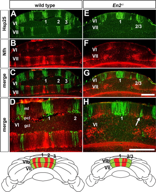
Hsp25 and Nfh molecular coding are altered in unison in the cerebellum of En2−/− mutant mice. A, Hsp25 expression reveals five ML stripes in lobules VI/VII of wild-type mice as seen on coronal cut tissue sections (#1 is the midline stripe and #2 and #3 flank both sides of the vermis midline). B–D, Hsp25 and Nfh are expressed in complementary ML stripes in lobules VI/VII of wild-type mice. E, The number of Hsp25 stripes is reduced from five to only three in lobules VI/VII of En2−/− mutants (#2/3 indicates the fused lateral stripes). F–H, Hsp25 and Nfh expression patterns are disrupted in lobules VI/VII of En2−/− mutants, but their complementary relationship is preserved. D and H are higher power images of the midline stripes shown in C and G. The arrow in H points to a patch of Purkinje cells that ectopically expresses Hsp25. The whole-mount schematics illustrate the complementary patterns of Hsp25 and Nfh in the CZ of wild-type and En2−/− mice. Scale bars: (in G), 500 μm (applies to A–C and E–G); (in H) 500 μm (applies to D).